Ultrasound Imaging is Vital in Diagnosing Chronic Venous Insufficiency
The term Chronic Venous Insufficiency (CVI) describes a condition that affects the venous system of the lower extremities with venous hypertension causing various pathologies including pain, swelling, edema, skin changes, and ulcerations.
Although the term CVI is often used to exclude uncomplicated varicose veins, varicose veins have incompetent valves with increased venous pressure leading to progressive dilation and twisting. We use the term CVI to represent the full spectrum of manifestations of chronic venous disease.
The Venous Ultrasound Exam
At the Center for Vein Restoration, the diagnosis of Chronic Venous Insufficiency is made via patient history and a physical examination with the assistance of noninvasive testing. A portion of the physical exam entails inspection and palpation of the surface of the skin to detect irregularities or bulges. The calf muscle is consistently assessed and measured for indications of fullness or increased girth due to long-standing edema.
Palpation may also reveal tenderness of the dilated veins. Active or healed ulcers are seen with more advanced disease.
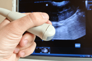
There is a broad differential for the common presenting complaint with CVI. As a result, noninvasive ultrasound imaging plays an important role in diagnosing and guiding CVI treatment. Ultrasound imaging, also called ultrasound scanning or sonography, involves exposing part of a patient’s body to high-frequency sound waves to produce pictures of the inside of the body. Unlike x-rays, ultrasound exams do not use ionizing radiation.
Ultrasound imaging helps us to diagnose and treat medical conditions because they can show the structure and movement of the body's internal organs, as well as blood flowing through blood vessels. A venous ultrasound provides us with pictures of the veins throughout the body that carry blood back to the heart.
This painless noninvasive exam measures the function of the vein valves that are critical to venous function, and can also identify vein blockages. In fact, compared to venography, which requires injecting contrast material into a vein, venous ultrasound is nearly as accurate for detecting blood clots in the calf and almost fully as accurate in finding clots in veins of the thigh.
A Doppler ultrasound may also be part of a venous ultrasound examination. This special ultrasound technique evaluates blood as it flows through a blood vessel, including the major arteries and veins, and the images can help us to see and evaluate blockages to blood flow, such as clots, or the narrowing of blood vessels.
While many patients present with large, clinically obvious bulging varicose veins, other individuals may have significant "silent" reflux (abnormal direction of blood flow) in diseased veins, which can only be detected by Doppler vein mapping a more detailed ultrasound evaluation of the abnormal veins of the legs which illustrates the path of the blood flowing through the abnormal veins and where the trouble begins.
Careful mapping of the lower extremity venous system aids us in determining the optimal treatment plan to meet each patient’s specific needs.